Dermapace System To Treat Diabetic Foot Ulcers

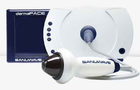
Dermapace System, the first shock wave device intended to treat diabetic foot ulcers.

“Diabetes is the leading cause of lower limb amputations,” said Binita Ashar, M.D., director of the division of surgical devices in FDA’s Center for Devices and Radiological Health. “The FDA is dedicated to making technologies available that can help improve the quality of life for those with chronic diseases. Additional options for successfully treating and healing ulcer wounds may help prevent lower limb amputations.”

An estimated 30.3 million people in the United States have been diagnosed with diabetes, according to the Centers for Disease Control and Prevention. Diabetes damages blood vessels and nerves, particularly in the feet, and can lead to severe infections that are difficult to treat. About 25 percent of people with diabetes will experience a foot ulcer in their lifetime. Amputation is sometimes necessary when circulation is so poor that a foot ulcer fails to heal or when treatment fails to stop the spread of an infection.

The Dermapace System is intended to be used in the treatment of chronic, full-thickness diabetic foot ulcers with wound areas measuring no larger than 16 cm2 (about the size of a soda can top) which extend through the epidermis, dermis, tendon, or capsule, but without bone exposure. The Dermapace System is an external (extracorporeal) shock wave system that uses pulses of energy, similar to sound waves, to mechanically stimulate the wound. The device is intended for adult patients (22 years and older), presenting with diabetic foot ulcers lasting for more than 30 days, and should be used along with standard diabetic ulcer care.

The FDA reviewed clinical data from two multi-center, randomized, double-blind studies with a total of 336 diabetic patients receiving either usual care, which includes wet-to-dry dressings or debridement (removal of damaged tissue) as needed, plus the Dermapace System shock wave therapy or usual care plus non-working (sham) shock wave therapy. Both patient groups included those with poorly controlled and well-controlled blood glucose levels.

The patients who had between one and seven treatments with the Dermapace System showed an increase in wound healing at 24 weeks with a 44 percent wound closure rate. Those patients treated with the sham shock wave therapy showed a 30 percent wound closure rate during the same time period.

The most common side effects observed were pain during application of the device, local bruising and numbness, migraines, nausea, fainting, wound infection, infection beyond the wound (cellulitis, osteomyelitis) and fever.

Cooling Cap to Reduce Hair Loss During Chemotherapy

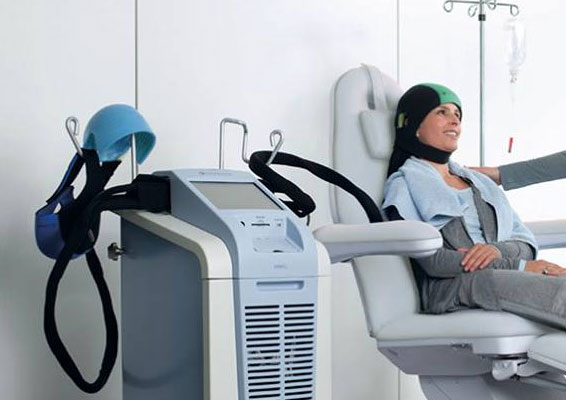
U.S. Food and Drug Administration cleared the expanded use of a cooling cap, DigniCap Cooling System, to reduce hair loss (alopecia) during chemotherapy. This is the first cooling cap cleared by the agency for use in cancer patients with solid tumors.

“We are pleased to expand the use of this product for cancer patients with solid tumors to potentially minimize chemotherapy-induced hair loss,” said Binita Ashar, M.D., director, Division of Surgical Devices, in the FDA’s Center for Devices and Radiological Health. “Managing the side effects of chemotherapy is a critical component to overall health and quality of life.”

Hair loss is a common side effect of certain types of chemotherapy and is commonly associated with the treatment of most solid tumor cancer. Hair may fall out entirely, gradually, in sections, or may become thin. Hair loss due to cancer treatment is usually temporary, but minimizing or relieving these kinds of side effects are considered important to overall treatment.

The DigniCap Cooling System is indicated to reduce the frequency and severity of hair loss during chemotherapy in solid tumor cancer patients in which alopecia-inducing chemotherapeutic agents and doses are used. It is a computer-controlled system used during treatment. A cap is worn on the head and circulates liquid to a cap to cool the scalp during chemotherapy treatment. The cap is covered by a second cap made from neoprene, a type of rubber that holds the cooling cap in place and acts as an insulation cover to prevent loss of cooling.

The cooling is intended to constrict blood vessels in the scalp, which reduces the amount of chemotherapy that reaches cells in the hair follicles. The cold temperature also decreases the activity of the hair follicles and slows down cell division, making them less affected by chemotherapy. The combined actions are thought to reduce the effect chemotherapy has on the cells, which may reduce hair loss. DigniCap may not work with some chemotherapy regimens.

The device is contraindicated for pediatric patients, patients with certain cancers and patient undergoing specific chemotherapy treatments. Additionally, DigniCap may not be appropriate for patients with cold sensitivity or susceptibility to cold-related injuries.

The most common side effects of the cooling system include cold-induced headaches and neck and shoulder discomfort, chills and pain associated with wearing the cooling cap for an extended period of time. The risk of the chemotherapy drug missing an isolated grouping of the cancer cells in the scalp because of the cooling cap is rare.

iGlucose Glucometer with Built-In Cellular Connectivity

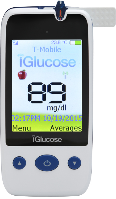
Smart Meter Corporation, a New York firm, won both FDA clearance and European CE Mark approval for its iGlucose system, a blood glucose meter that features built-in cellular connectivity. Diabetics using the iGlucose will have the convenience of  seeing their readings automatically uploaded to the company’s web portal, from where their doctors and caretakers can check up on how things are going.

Blood glucose readings are shared with the cloud-based iGlucose Web Portal as soon as they’re taken, and from just about anywhere there is a cellular signal. Lifetime use of the cellular network is included with the device, so there is no subscription to pay for every month.

The glucometer needs only .75 µl of blood and provides results in just a few seconds and the company says its test strips are price competitive with popular strips on the market now.

When unusually high or low sugar levels are detected, the iGlucose Web Portal can be made to automatically send out alerts via email or text messages to the family and clinical team of the patient, helping to address issues quicker and before more serious consequences kick in.

An interesting feature of the iGlucose system is that it comes with an API (Application Program Interface), allowing outside developers to integrate their applications with the iGlucose. We’re glad to see this is becoming more common among medical device makers, helping to facilitate a more interconnected world of medical information.

Myoscience iovera System Cleared for Treating Painful Osteoarthritic Knees

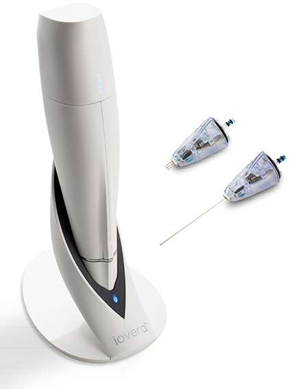
Myoscience, a company based in Fremont, California won FDA clearance for its iovera cryoneurolysis system to be used for symptom and pain relief in the knees arising from osteoarthritis. The company’s so-called Focused Cold Therapy delivery system pumps pressurized liquid nitrous oxide (N2O) to the treatment tip, where it is made to turn to a gas and become very cold. The phase change happens in tight location, creating a small and precise treatment area that can be used to create a nerve block that stops the transmission of pain.

Treatments are quite fast and result in nearly instantaneous pain reduction thanks to a disruption in the functionality of nerve axons. This happens without damaging the surrounding tissue that surrounds the nerves.

Here’s a bit about the study that led to the latest clearance:

The FDA clearance was based on a recent prospective, multi-center, sham-controlled, randomized, double-blind study of 180 subjects. The study concluded that patients treated with the iovera° device reported statistically significant greater reduction in pain and improvement in symptoms (p=0.001) when compared to patients who received the sham treatment(3). Also, patients who received the iovera° treatment reported pain relief lasting up to 90 days, accompanied by reduced stiffness and improved physical function.

Standard test may miss urinary infection in symptomatic women

New research from Belgium suggests that the standard culture test for bacteria may return a negative result even though the patient tested actually has a urinary tract infection. The study compared women with symptoms of urinary infection – such as pain during urination and feeling an urgent or frequent need to urinate – with non-symptomatic women. With the help of a more sensitive test, it found that nearly all the symptomatic women with a negative standard test result actually did have an infection.

Lead author Dr. Stefan Heytens, a researcher at Ghent University and a practicing GP, says that their findings support the idea that, for women with symptoms of an uncomplicated UTI, there is no need to carry out a standard culture test.


Standard test is negative for many symptomatic women

Dr. Heytens and colleagues note that around 20 to 30 percent of women with symptoms of UTI who have the standard culture test have a negative result.


Negative standard test ‘assumed to mean no UTI’

If the result of the standard test is positive, then the patient may be offered a course of antibiotics. These can include fosfomycin, nitrofurantoin, or trimethoprim.

However, the researchers say that many doctors assume that if the test result is negative, then the patient does not have a UTI.

They note that there has also been a tendency, in the past, to consider women who fall into this category as having unexplained ‘urethral syndrome’, and that the cause is likely to be psychosomatic.

For their study, Dr. Heytens and colleagues compared urine samples from 220 women who went to see their doctor because they had symptoms of UTI, with those of 86 healthy women with no such symptoms.

The urine samples underwent the standard culture test and were also tested with a more sensitive method called quantitative polymerase chain reaction (qPCR), which can detect minute amounts of bacterial DNA known to cause UTIs, including Escherichia coli and Staphylococcus saprophyticus.

‘If a woman has symptoms, she probably has a UTI’

The researchers found that the standard test detected bacteria in 80.9 percent of urine samples from the symptomatic women.

However, the qPCR test detected E. coli in 95.9 percent of those samples, and S. saphrophyticus in 8.6 percent.

When the two qPCR test results were combined, they showed that 98.2 percent of the symptomatic women had an infection.

In the symptom-free, healthy volunteers, the standard test found evidence of E. coli in 10.5 percent of them, and the qPCR test found E. coli in 11.6 percent.

Dr. Heytens says that the more sensitive qPCR test found evidence of E. coli in the urine samples of nearly all the women presenting with UTI symptoms, even when their standard test returned a negative result.

“This suggests that if a woman has these symptoms, she probably does have a UTI,” he suggests, and concludes:

“Our findings support previous research which indicates that traditional testing may not be helpful in uncomplicated UTIs. However, traditional urine culture tests may still have a role to play if treatment fails or if there are signs and symptoms of a more complicated UTI.”

However, he also points out that they still do not know if antibiotics are of benefit to all women who present with UTI symptoms.

The study – by researchers from Ghent University and Ghent University Hospital in Belgium – is published in the journal Clinical Microbiology and Infection.

3D Printed Skin for Transplants

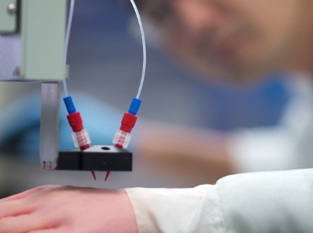
A collaboration of Spanish researchers from Universidad Carlos III de Madrid, the Center for Energy, Environmental and Technological Research, Hospital General Universitario Gregorio Marañón, and BioDan Group, a bioengineering company, have reportedly developed a 3D printer that produces human skin ready for transplantation. Besides obvious applications such as producing replacement skin for burn victims, it can be used to test drugs, new therapies for skin conditions, and for evaluating the safety and effectiveness of cosmetic products. The researchers believe it will function about as well as natural skin and it is already being evaluated by European regulatory agencies for clinical use.

The printed skin has both dermis and epidermis layers, as well as the stratum corneum, the exterior layer that is made of dead skin cells. The dermis is made of fibroblasts and generate collagen, making the material extremely similar to natural skin in terms of pliability and strength.

The technology relied on developing and learning how to use a set of biological inks, consisting of things like cells and proteins, so when they’re brought together they work as intended. “Knowing how to mix the biological components, in what conditions to work with them so that the cells don’t deteriorate, and how to correctly deposit the product is critical to the system,” said Juan Francisco del Cañizo, of the Hospital General Universitario Gregorio Marañón and Universidad Complutense de Madrid.

“This method of bioprinting allows skin to be generated in a standardized, automated way, and the process is less expensive than manual production,” said Alfredo Brisac, CEO of BioDan Group, in a statement.

1 in 4 men with suspected prostate cancer could avoid unnecessary biopsy if given an MRI scan first

Giving men with suspected prostate cancer an MRI scan could improve diagnosis and save those who do not have aggressive cancers from having an unnecessary biopsy, according to a study published in The Lancet.


The study estimates that adding the extra test could help one in four (27%) men avoid an unnecessary biopsy and reduce the number of men who are over-diagnosed – diagnosed with a cancer that does not go on to cause any harm during their lifetime – by 5%.


Typically, men undergo a biopsy of their prostate if they experience symptoms of prostate cancer or have a prostate specific antigen (PSA) test showing high levels of the PSA protein in their blood. Each year, over 100,000 prostate biopsies are carried out in the UK and one million are conducted in Europe. However, the PSA test is not always accurate, which means that many men undergo unnecessary biopsies.


“Prostate cancer has aggressive and harmless forms. Our current biopsy test can be inaccurate because the tissue samples are taken at random. This means it cannot confirm whether a cancer is aggressive or not and can miss aggressive cancers that are actually there. Because of this some men with no cancer or harmless cancers are sometimes given the wrong diagnosis and are then treated even though this offers no survival benefit and can often cause side effects. On top of these errors in diagnosis, the current biopsy test can cause side effects such as bleeding, pain and serious infections.” said lead author, Dr Hashim Ahmed, UCL, UK. [1]


Multi-parametric MRI (MP-MRI) scans provide information about the cancer’s size, how densely packed its cells are and how well connected to the bloodstream it is, so could help differentiate between aggressive and harmless cancers.


In this study, 576 men with suspected prostate cancer were given an MP-MRI scan followed by two types of biopsy in 11 NHS hospitals. Firstly, they underwent a template prostate mapping (TPM) biopsy, which was used as a control to compare the accuracy of the MP-MRI and standard biopsy against. The second biopsy was the standard transrectal ultrasound-guided (TRUS) biopsy – the most commonly used biopsy for diagnosing prostate cancer.


The TPM biopsy found that less than half of the men in the study (40%) had aggressive cancer.


Of these, the MP-MRI scan correctly diagnosed almost all of the aggressive cancers (93%), whereas the TRUS biopsy correctly diagnosed only half (48%). Further, for men who had a negative MP-MRI scan, nine out of 10 (89%) had either no cancer or a harmless cancer.


Because of this, the researchers suggest that MP-MRI could be used before TRUS biopsy to identify those who have harmless cancers and do not need a biopsy immediately. This group could instead continue to be monitored by their doctors, while those thought to have aggressive cancers could then have their MP-MRI scan result confirmed by the TRUS biopsy. Overall, this would reduce over-diagnosis while improving detection of aggressive cancers.


“Our results show that MP-MRI should be used before biopsy. Our study found that using the two tests could reduce over-diagnosis of harmless cancers by 5%, prevent one in four men having an unnecessary biopsy, and improve the detection of aggressive cancers from 48% to 93%,” said Dr Hashim Ahmed. “While combining the two tests gives better results than biopsy alone, this is still not 100% accurate so it would be important that men would still be monitored after their MP-MRI scan. Biopsies will still be needed if an MP-MRI scan shows suspected cancer too, but the scan could help to guide the biopsy so that fewer and better biopsies are taken.” 1


During the study there were 44 serious adverse events, with eight cases of sepsis caused by a urinary tract infection and 58 cases of urinary retention. These were a result of the biopsies rather than the MP-MRI scan, and are symptoms commonly seen in the clinic as a result of the standard biopsy.


Limitations of the study include that giving the TPM biopsy before the TRUS biopsy may have caused swelling and changes to the prostate tissue which could affect the accuracy of the TRUS biopsy. In addition, more research is needed into the cost-effectiveness of this approach, how it affects hospital capacity and ensuring there are enough radiologists to conduct the MP-MRI in the NHS.


The study was funded by the UK Department of Health, National Institute for Health Research, The University College London Hospitals Biomedical Research Centre and the Royal Marsden and Institute of Cancer Research Biomedical Research Centre. It was conducted by scientists from UCL, UCLH NHS Foundation Trust, the Royal Marsden Hospital, the MRC Clinical Trials Unit at UCL, University of York and Hampshire Hospitals NHS Foundation Trust.


Article: Diagnostic accuracy of multi-parametric MRI and TRUS biopsy in prostate cancer (PROMIS): a paired validating confirmatory study, Hashim U Ahmed, FRC et al., The Lancet, doi: 10.1016/S0140-6736(16)32401-1, published 19 January 2017.

Eko Core Digital Stethoscope

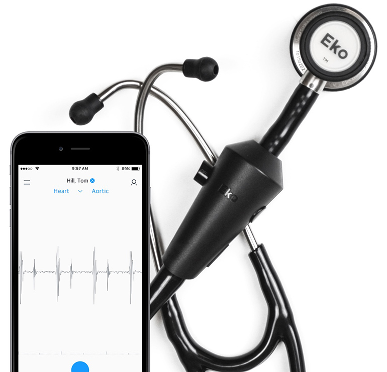
Eko Core, the digital stethoscope from Eko Devices, a Berkeley, CA firm. It’s much more than a high-fidelity amplified stethoscope. You can store and share auscultations, as well as analyze and annotate them using a paired app, which wirelessly connects to the Eko Core. You can even listen to recordings shared by other clinicians through the Eko Core, so you can hear exactly what they heard when they conducted the exam.

Everything can be stored to an electronic medical record (EMR), just like a radiologist would save patient CT scans, and these can be compared during future exams. The data can even be live-streamed for tele-medicine applications, such as allowing cardiology specialists to help to diagnose patients in remote and understaffed clinics. All the sharing and streaming is done using HIPAA-compliant software, allowing it to be used and integrated into existing clinical practices and their IT systems.

When you get your stethoscope, you download the app and connect the stethoscope to your iPhone, iPad, or Android device via Bluetooth.

When you perform an auscultation exam, keep your iPhone handy: the software will automatically navigate you to record, store, or share the sounds. You can annotate and attribute recorded sounds to specific regions of the chest or back, say aortic valve region for heart sounds, or upper left for lungs. (Attributing sounds to specific locations is optional. You can just record the sounds on their own if you wish to.)

The sharing part is HIPAA compliant, and you can share the recordings with colleagues or save it to an EMR. You can also receive recordings from other clinicians and hear them right there in the stethoscope. So the stethoscope is not only a recording device, but also a playback device.

Medical Robotics Shows Off Its New Versius System

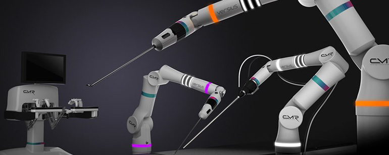
Cambridge Medical Robotics, a UK firm, is revealing its Versius robotic surgery system. The system consists of modular robotic arms, any number of which can be used depending on a procedure. The arms can have a camera or any one of the dozen or so tools attached, and they can be quickly swapped for other tools as necessary.

Each of the arms can be placed around the patient table or even hung from above to save valuable space. The surgeon wears a pair of 3D glasses and operates by looking at a monitor instead of peering into a scope common on existing systems. This can help improve ergonomics and allow the surgeon to see and interact easier with clinicians managing the patient and the robot. The robot is operated using a controller similar to video game joysticks and the system delivers haptic feedback from the instrument to the controller, so the surgeon can actually feel the anatomy being worked on.

Unlike existing robotic surgical systems, the Versius can work with instruments requiring only a 5 mm incision. Typically the smallest instrument sizes on robotic systems is 8 mm, and unlike 5 mm incisions these typically require suturing and maintenance.

The company has already performed a number of studies, including on cadavers, and is compiling responses and data from 32 surgeons that has already used the Versius system. The firm hopes to receive the CE Mark in Europe in 2018 and FDA regulatory green light shortly thereafter.

SnooZeal Prevents Snoring by Training Tongue During Daytime

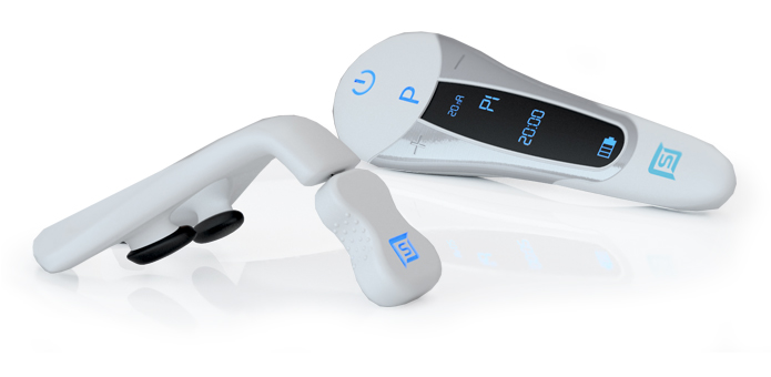
The SnooZeal product consists of a mouth piece that places electrodes above and below the tongue, a control unit that connects to the mouthpiece, a remote control, and a smartphone app. It works by electrically stimulating the tongue to give it a workout and keep it from completely relaxing and collapsing during the night. Snoozeal Inc. is a company out of Seattle, Washington that won the European CE Mark for its snoring prevention device.

The SnooZeal is not actually used during sleep, but indicated to be placed in the mouth twice a day for a period of six weeks. This essentially physically trains the tongue muscle and helps to keep it at least partially contracted even at night.

The device can be controlled either via the remote or through the accompanying smartphone app.

Results from multiple previous clinical trials have proven that muscle activity can be improved with this electrical stimulation technology principle.

Several studies have shown that electrical stimulation of the tongue can reduce snoring and sleep-related obstructions.

SnooZeal is the first patented product that is used entirely in the mouth and that directly stimulates the tongue muscle in order to address the root cause of snoring.