Low-Dose Aspirin Linked to Reduced Risk of Breast Cancer

Protective link particularly significant for hormone receptor-positive/HER2-negative cancer

Regularly taking low-dose aspirin appears to protect women from hormone receptor-positive/human epidermal growth factor receptor 2 (HER2)-negative breast cancer, according to a study published online May 1 in Breast Cancer Research.

Christina Clarke, Ph.D., M.P.H., of the Cancer Prevention Institute of California in Fremont, and colleagues looked at the medication use of women enrolled in the ongoing California Teachers Study. That trial, begun in 1995, recruited 133,479 active and retired women teachers, administrators, and other public school professionals. In 2005, 57,164 participants answered questions about their use of aspirin and other medications, family history of cancer, use of hormone therapy, alcohol use, exercise, height, and weight. By 2013, 1,457 had developed invasive breast cancer.

Overall, the researchers found that use of low-dose aspirin at least three times a week was associated with a 16 percent reduced risk of breast cancer. But the more significant finding was the risk reduction for developing hormone receptor-positive/HER2-negative cancer. The researchers found a protective link with use of low-dose aspirin, but not with regular-dose aspirin or other nonsteroidal anti-inflammatory drugs such as ibuprofen or acetaminophen.

“Our observation of reduced risk of breast cancer, among participants who took three or more tablets of low-dose aspirin weekly, is consistent with other reports looking at aspirin without differentiation by dose. This is the first report to suggest that the reduction in risk occurs for low-dose aspirin and not for regular-dose aspirin and only among women with the hormone receptor-positive/HER2-negative subtype,” the authors write. “This preliminary study builds on previous knowledge and further supports the need for formal cancer chemoprevention studies of low-dose aspirin.”

Standard test may miss urinary infection in symptomatic women

New research from Belgium suggests that the standard culture test for bacteria may return a negative result even though the patient tested actually has a urinary tract infection. The study compared women with symptoms of urinary infection – such as pain during urination and feeling an urgent or frequent need to urinate – with non-symptomatic women. With the help of a more sensitive test, it found that nearly all the symptomatic women with a negative standard test result actually did have an infection.

Lead author Dr. Stefan Heytens, a researcher at Ghent University and a practicing GP, says that their findings support the idea that, for women with symptoms of an uncomplicated UTI, there is no need to carry out a standard culture test.


Standard test is negative for many symptomatic women

Dr. Heytens and colleagues note that around 20 to 30 percent of women with symptoms of UTI who have the standard culture test have a negative result.


Negative standard test ‘assumed to mean no UTI’

If the result of the standard test is positive, then the patient may be offered a course of antibiotics. These can include fosfomycin, nitrofurantoin, or trimethoprim.

However, the researchers say that many doctors assume that if the test result is negative, then the patient does not have a UTI.

They note that there has also been a tendency, in the past, to consider women who fall into this category as having unexplained ‘urethral syndrome’, and that the cause is likely to be psychosomatic.

For their study, Dr. Heytens and colleagues compared urine samples from 220 women who went to see their doctor because they had symptoms of UTI, with those of 86 healthy women with no such symptoms.

The urine samples underwent the standard culture test and were also tested with a more sensitive method called quantitative polymerase chain reaction (qPCR), which can detect minute amounts of bacterial DNA known to cause UTIs, including Escherichia coli and Staphylococcus saprophyticus.

‘If a woman has symptoms, she probably has a UTI’

The researchers found that the standard test detected bacteria in 80.9 percent of urine samples from the symptomatic women.

However, the qPCR test detected E. coli in 95.9 percent of those samples, and S. saphrophyticus in 8.6 percent.

When the two qPCR test results were combined, they showed that 98.2 percent of the symptomatic women had an infection.

In the symptom-free, healthy volunteers, the standard test found evidence of E. coli in 10.5 percent of them, and the qPCR test found E. coli in 11.6 percent.

Dr. Heytens says that the more sensitive qPCR test found evidence of E. coli in the urine samples of nearly all the women presenting with UTI symptoms, even when their standard test returned a negative result.

“This suggests that if a woman has these symptoms, she probably does have a UTI,” he suggests, and concludes:

“Our findings support previous research which indicates that traditional testing may not be helpful in uncomplicated UTIs. However, traditional urine culture tests may still have a role to play if treatment fails or if there are signs and symptoms of a more complicated UTI.”

However, he also points out that they still do not know if antibiotics are of benefit to all women who present with UTI symptoms.

The study – by researchers from Ghent University and Ghent University Hospital in Belgium – is published in the journal Clinical Microbiology and Infection.

Even Short-term Oral Steroids Carry Serious Risk

The millions of Americans prescribed short-term oral corticosteroids are taking a dose of risk along with their medication, according to a cohort study of more than 1.5 million adults.

Within 30 days of initiating these drugs, even at relatively low doses, users had a nearly twofold increased risk for fracture, a threefold increased risk for venous thromboembolism, and a fivefold increased risk for sepsis.

“Greater attention to initiating prescriptions of these drugs and monitoring for adverse events may potentially improve patient safety,” write Akbar K. Waljee, MD, an assistant professor of gastroenterology at the University of Michigan in Ann Arbor, and colleagues. They present their findings in an article published April 12 in the BMJ.

They found that more than one in five adults included in the Clinformatics DataMart, a large national database of commercial insurance claims, received prescriptions for short-term oral corticosteroids during the 3-year study, which ran from January 1, 2012, to December 31, 2014.

Although corticosteroids are among the most common cause for hospitalization for drug-related adverse events, and various specialties have long focused on optimizing their long-term use, the short-term risks associated with the drugs have been less carefully studied.

“Although physicians focus on the long-term consequences of steroids, they don’t tend to think about potential risks from short-term use,” said Dr Waljee in a university news release. “We see a clear signal of higher rates of these three serious events within 30 days of filling a prescription. We need to understand that steroids do have a real risk and that we may use them more than we really need to. This is so important because of how often these drugs are used.”

Of 1,548,945 adults aged 18 to 64 years included in the database, 327,452 (21.1%) received at least one outpatient prescription for short-term oral corticosteroids (30 or fewer days). The mean age of users was 45.5 years (standard deviation [SD], 11.6 years) compared with 44.1 years (SD, 12.2 years) for nonusers (P < .001). The median duration of use was 6 days (interquartile range, 6 – 12 days).

The six most common indications for the drugs were upper respiratory tract infections, spinal conditions, intervertebral disc disorders, allergies, bronchitis, and nonbronchitic lower respiratory tract disorders. Together, those indications accounted for about half of all prescriptions. The two most common physician specialties prescribing short-term oral corticosteroids were family medicine and general internal medicine.

Nearly half (46.9%) of recipients were prescribed a 6-day prepackaged methylprednisolone “dosepak,” which tapers the dose from highest to lowest. Dr Waljee noted in the news release that although individual oral steroid pills can cost less than a dollar each for a 7-day course, the prepackaged version may cost several times that and often initiates therapy with a higher high dose than may be necessary.

Use was more frequent among older patients, women, and white adults, with significant regional variation (all P < .001).

Within 30 days of drug initiation, there was an increase in incidence rate of the following: sepsis, with a rate ratio of 5.30 (95% CI, 3.80 – 7.41); venous thromboembolism, with a rate ratio of 3.33 (95% CI, 2.78 – 3.99); and fracture, with a rate ratio of 1.87 (95% CI, 1.69 – 2.07).

The increased risk persisted at prednisone equivalent doses of less than 20 mg/day, with an incidence rate ratio of 4.02 for sepsis, 3.61 for venous thromboembolism, and 1.83 for fracture (all P < .001).

Rate ratios decreased during the following subsequent 31 to 90 days, however.

Although rare, hospitalizations were also more frequent in users than nonusers, with 0.05% of users admitted for sepsis compared with 0.02% of nonusers. For blood clots, the admission rate was 0.14% versus 0.09%, and for fractures, it was 0.51% vs 0.39%.

Dr Waljee and associates found it significant that the most frequent corticosteroid prescribers were not rheumatologists or other subspecialists experienced in treating inflammatory conditions long-term. “A substantial challenge to improving use of oral corticosteroids will be the diverse set of conditions and types of providers who administer these drugs in brief courses,” they write. “This raises the need for early general medical education of clinicians about the potential risks of oral corticosteroids and the evidence basis for their use, given that use may not be specific to a particular disease or specialty.”

On the basis of these findings, Dr Waljee recommended prescribing the smallest possible amount of corticosteroids for treating the condition in question. “If there are alternatives to steroids, we should be use those when possible,” he said in the release. “Steroids may work faster, but they aren’t as risk-free as you might think.”

BMJ. 2017;357:j1415

Antidepressants in Pregnancy: No Link to Autism, ADHD

Use of antidepressants before and during pregnancy does not cause autism, or attention-deficit/hyperactivity disorder (ADHD) new research shows.

Three studies demonstrate that antidepressant use in pregnant women is likely not responsible for autistic spectrum disorders (ASDs) in children and that the association found in previous studies was likely due to confounding factors.

A team of researchers headed by Simone N. Vigod, MD, of Women’s College Hospital, Toronto, Canada, evaluated the association between gestational serotonergic antidepressant exposure and childhood ASD in 35,906 births.

In the 2837 pregnancies (7.9%) involving exposure to antidepressants, 2% of children were diagnosed with ASD, a percantage that was higher than in the children who had not been exposed. However, once the researchers adjusted for confounding factors, the difference was no longer significant. Nor was it significant when exposed children were compared with unexposed siblings.

“We will never be able to say for certain that a medication has zero risks,” Dr Vigod told Medscape Medical News. “But these data, together with the findings of the other studies published this week, are reassuring and suggest that antidepressants during pregnancy are reasonably low risk.”

In another study, Brian M D’Onofrio, PhD, of Indiana University, Bloomington, and colleagues focused on the confounding factors between first-trimester antidepressant exposure and birth and neurodevelopmental problems.

The researchers analyzed a database of more than 1.5 million Swedish offspring born between 1992 and 2012 for whom follow-up was available through 2013. Of these, 1.4% (n = 22,544) were born to mothers who self-reported antidepressant use during their first trimester.

The researchers used sophisticated statistical methods to account for a variety of confounding factors. After controlling for these factors, they found that first-trimester antidepressant exposure, as compared to nonexposure, was associated with a “small increased risk of preterm birth,” but not with increased risk for having small size for gestational age, ASD, or ADHD.

“Our study suggests that it is not the medication use during pregnancy per se that explains why children of mothers who use these medications have more problems than children whose mothers do not,” Dr D’Onofrio told.

The studies were published in the April 18 issue of JAMA.

The third article is a meta-analysis conducted by Florence Gressier, MD, PhD, of the Bicêtre University Hospital, Le Kremlin-Bicêtre, France, and colleagues. The researchers analyzed 10 studies with “inconsistent results” regarding the association of ASD with fetal exposure to antidepressants for each trimester and during the preconception period.

Although the researchers found a significant association between increased ASD risk and maternal use of antidepressants during pregnancy, the association was more consistent during the preconception period than during each trimester. Moreover, the association was weaker when the researchers controlled for past maternal mental illness.

“Previously reported differences between exposed and unexposed children may come from statistical fluctuations, different criteria of evaluation, and different modelings,” Dr Gressier told.

This study was published in the April 17 issue of JAMA Pediatrics.

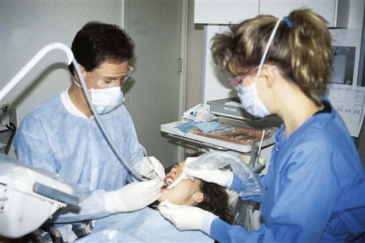
Preventing Disease Transmission in Dental Settings

Infection Transmission in Dental Healthcare

Reports of transmission of infectious agents between patients and dental healthcare personnel (DHCP) in dental settings are rare. However, a recent Centers for Disease Control and Prevention (CDC) article in the Journal of the American Dental Association[1] identified three published reports describing the transmission of hepatitis B virus and hepatitis C virus in dental settings since 2003. In addition, the Morbidity and Mortality Weekly Report[2]—published April 8, 2016—described a 2015 outbreak of Mycobacterium abscessus infection at a pediatric dentistry practice.

In most cases, investigators have failed to link a specific lapse of infection prevention and control practice with a particular transmission. However, reported breakdowns in basic infection prevention practices included unsafe injection practices, failure to heat-sterilize dental handpieces between patients, failure to monitor (eg, conduct spore testing of) autoclaves, and failure to maintain dental unit waterlines. These reports highlight the need to improve understanding of and compliance with current infection prevention recommendations.

Key information and recommendations relevant to the above identified breaches include:

  • When administering local anesthesia, use needles and anesthetic cartridges for one patient only and clean and heat-sterilize the dental cartridge syringe between patients.
  • If multidose vials are used (such as for conscious sedation), dedicate multidose vials to a single patient whenever possible.
  • If multidose vials must be used for more than one patient, restrict them to a centralized medication area and do not allow them to enter the treatment area.
  • If a multidose vial enters the immediate patient treatment area, it should be dedicated for single-patient use and discarded immediately after use.
  • Date multidose vials when first opened and discard within 28 days, unless the manufacturer specifies a shorter or longer date for discarding that opened vial.
  • Clean and heat-sterilize all handpieces (high-speed and low-speed) and attachments that attach to air and waterlines (including motors) between patients unless they are single-use, disposable items. For handpieces that do not connect to air and waterlines, use US Food and Drug Administration–cleared devices and follow the validated manufacturer’s instructions for reprocessing.
  • Monitor sterilizers at least weekly by using a biological indicator with a matching control (ie, biological indicator and control from same lot number). Results of biological monitoring should be recorded and sterilization monitoring records (mechanical, chemical, and biological) retained long enough to comply with state and local regulations.
  • Ensure that all dental units use systems that treat water to meet drinking water standards (ie, ≤ 500 colony forming units/mL of heterotrophic water bacteria). Consult with the dental unit manufacturer for appropriate water maintenance methods and recommendations for monitoring dental water quality.

CDC Division of Oral Health

ArmonAir RespiClick (fluticasone propionate) for Maintenance Treatment of Asthma

Teva Pharmaceutical Industries Ltd., (NYSE and TASE: TEVA) today announced that the U.S. Food and Drug Administration (FDA) has approved ArmonAir RespiClick (fluticasone propionate inhalation powder) for adolescent and adult patients with asthma. The medication is delivered via Teva’s RespiClick® breath-activated, multi-dose dry powder inhaler (MDPI) which is used with other approved medicines in Teva’s respiratory product portfolio.

ArmonAir RespiClick is an inhaled corticosteroid (ICS) containing the same active ingredient as Flovent, and is indicated for the maintenance treatment of asthma as prophylactic therapy in patients 12 years and older.

ArmonAir RespiClick is expected to become available to patients in the U.S., by prescription, later this year. The approved strengths of ArmonAir RespiClick are: 55 mcg, 113 mcg, and 232 mcg administered as one inhalation twice daily.

The FDA approval of ArmonAir RespiClick is supported by data from Teva’s clinical development program, including three Phase III trials which evaluated the efficacy and safety of the treatment in adolescent and adult patients with asthma. In the two double-blind studies, the therapy showed clinically relevant and greater benefit compared with placebo in the improvement of lung function after 12 weeks of treatment as measured by Forced Expiratory Volume in one second (FEV1). In ArmonAir RespiClick clinical trials, the most common adverse reactions (incidence ≥3%) were nasopharyngitis, headache, cough, oral candidiasis and upper respiratory tract infection.

About ArmonAir RespiClick (Fluticasone Propionate) Inhalation Powder

ArmonAir RespiClick is indicated for the maintenance treatment of asthma as prophylactic therapy in patients 12 years of age and older.

Important Limitation of Use: ArmonAir RespiClick is NOT indicated for the relief of acute bronchospasm.

IMPORTANT SAFETY INFORMATION

Contraindications: ArmonAir RespiClick is contraindicated in:

  • the primary treatment of status asthmaticus or other acute episodes of asthma where intensive measures are required
  • patients with known severe hypersensitivity to milk proteins or known hypersensitivity to fluticasone propionate or any of the excipients


  • Local Effects: Oropharyngeal candidiasis has occurred in patients treated with ArmonAir RespiClick. Advise patients to rinse the mouth with water without swallowing following inhalation
  • Acute Asthma Episodes: ArmonAir RespiClick is not indicated for the relief of acute bronchospasm. An inhaled, short-acting beta2-agonist, not ArmonAir RespiClick, should be used to relieve acute symptoms such as shortness of breath
  • Immunosuppression: Patients on corticosteroids are at risk for potential worsening of existing tuberculosis; fungal, bacterial, viral, or parasitic infections; or ocular herpes simplex. A more serious or even fatal course of chickenpox or measles may occur in susceptible patients. Use with caution, if at all, in patients with the above because of the potential for worsening of these infections
  • Transferring Patients from Systemic Corticosteroid Therapy: Particular care is needed for patients who have been transferred from systemically active corticosteroids to inhaled corticosteroids because deaths due to adrenal insufficiency have occurred in patients with asthma during and after transfer from systemic corticosteroids to less systemically available inhaled corticosteroids. Slowly taper the dose of systemic corticosteroids if transferring patients to ArmonAir RespiClick
  • Hypercorticism and Adrenal Suppression: Because of the possibility of significant systemic absorption of inhaled corticosteroids, patients on ArmonAir RespiClick should be observed carefully for any evidence of systemic corticosteroid effects. If such effects occur, the dosage of ArmonAir RespiClick should be reduced slowly, consistent with accepted procedures for reducing systemic corticosteroids, and for management of asthma symptoms
  • Hypersensitivity Reactions, Including Anaphylaxis: Immediate hypersensitivity reactions (e.g., urticaria, angioedema, rash, bronchospasm, hypotension), including anaphylaxis, may occur after administration of ArmonAir RespiClick. Discontinue ArmonAir RespiClick if such reactions occur
  • Reduction in Bone Mineral Density (BMD): Decreases in BMD have been observed with long-term administration of inhaled corticosteroids. Patients with major risk factors for decreased bone mineral content, such as prolonged immobilization, family history of osteoporosis, or chronic use of drugs that can reduce bone mass (e.g., anticonvulsants, oral corticosteroids) should be monitored and treated with established standards of care Effect on Growth: Inhaled corticosteroids, including ArmonAir RespiClick, may cause a reduction in growth velocity when administered to pediatric patients. Monitor the growth of pediatric patients receiving ArmonAir RespiClick routinely (e.g., via stadiometry). Titrate to the lowest dosage that effectively controls symptoms
  • Glaucoma and Cataracts: Glaucoma, increased intraocular pressure, and cataracts have been reported in patients with asthma following the long-term administration of inhaled corticosteroids, including fluticasone propionate. Close monitoring is warranted in patients with a change in vision or with a history of increased intraocular pressure, glaucoma, and/or cataracts
  • Paradoxical Bronchospasm: Bronchospasm may occur with an immediate increase in wheezing after dosing and should be treated immediately with an inhaled, short-acting bronchodilator; ArmonAir RespiClick should be discontinued immediately and alternative therapy instituted
  • Drug Interactions with Strong Cytochrome P450 3A4 Inhibitors: The use of strong cytochrome P450 3A4 (CYP3A4) inhibitors (e.g., ritonavir, atazanavir, clarithromycin, indinavir, itraconazole, nefazodone, nelfinavir, saquinavir, ketoconazole, telithromycin) with ArmonAir RespiClick is not recommended because increased systemic corticosteroid adverse effects may occur
  • Eosinophilic Conditions and Churg-Strauss Syndrome: Systemic eosinophilic conditions, such as Churg-Strauss syndrome, may occur. These events usually, but not always, have been associated with the reduction and/or withdrawal of oral corticosteroids following the introduction of fluticasone propionate. Be alert to eosinophilia, vasculitic rash, worsening pulmonary symptoms, cardiac complications, and/or neuropathy
  • Adverse Reactions: Most common adverse reactions (≥3%) in patients taking ArmonAir RespiClick 55 mcg twice daily, 113 mcg twice daily, 232 mcg twice daily, and placebo, respectively, were nasopharyngitis (5.4%, 5.8%, 4.8%, 4.4%), upper respiratory tract infection (5.4%, 4.7%, 5.5%, 4.8%), oral candidiasis (3.1%, 2.9%, 4.8%, 0.7%), headache (1.6%, 7.3%, 4.8%, 4.4%), and cough (1.6%, 1.8%, 3.4%, 2.6%)
  • Drug Interactions: The use of strong cytochrome P450 3A4 (CYP3A4) inhibitors (e.g., ritonavir, atazanavir, clarithromycin, indinavir, itraconazole, nefazodone, nelfinavir, saquinavir, ketoconazole, telithromycin) with ArmonAir RespiClick is not recommended because increased systemic corticosteroid adverse effects may occur
  • Use in Specific Populations: Since fluticasone propionate is predominantly cleared by hepatic metabolism, impairment of liver function may lead to accumulation of fluticasone propionate in plasma. Patients with hepatic disease should be closely monitored


3D Printed Skin for Transplants

A collaboration of Spanish researchers from Universidad Carlos III de Madrid, the Center for Energy, Environmental and Technological Research, Hospital General Universitario Gregorio Marañón, and BioDan Group, a bioengineering company, have reportedly developed a 3D printer that produces human skin ready for transplantation. Besides obvious applications such as producing replacement skin for burn victims, it can be used to test drugs, new therapies for skin conditions, and for evaluating the safety and effectiveness of cosmetic products. The researchers believe it will function about as well as natural skin and it is already being evaluated by European regulatory agencies for clinical use.

The printed skin has both dermis and epidermis layers, as well as the stratum corneum, the exterior layer that is made of dead skin cells. The dermis is made of fibroblasts and generate collagen, making the material extremely similar to natural skin in terms of pliability and strength.

The technology relied on developing and learning how to use a set of biological inks, consisting of things like cells and proteins, so when they’re brought together they work as intended. “Knowing how to mix the biological components, in what conditions to work with them so that the cells don’t deteriorate, and how to correctly deposit the product is critical to the system,” said Juan Francisco del Cañizo, of the Hospital General Universitario Gregorio Marañón and Universidad Complutense de Madrid.

“This method of bioprinting allows skin to be generated in a standardized, automated way, and the process is less expensive than manual production,” said Alfredo Brisac, CEO of BioDan Group, in a statement.

1 in 4 men with suspected prostate cancer could avoid unnecessary biopsy if given an MRI scan first

Giving men with suspected prostate cancer an MRI scan could improve diagnosis and save those who do not have aggressive cancers from having an unnecessary biopsy, according to a study published in The Lancet.


The study estimates that adding the extra test could help one in four (27%) men avoid an unnecessary biopsy and reduce the number of men who are over-diagnosed – diagnosed with a cancer that does not go on to cause any harm during their lifetime – by 5%.


Typically, men undergo a biopsy of their prostate if they experience symptoms of prostate cancer or have a prostate specific antigen (PSA) test showing high levels of the PSA protein in their blood. Each year, over 100,000 prostate biopsies are carried out in the UK and one million are conducted in Europe. However, the PSA test is not always accurate, which means that many men undergo unnecessary biopsies.


“Prostate cancer has aggressive and harmless forms. Our current biopsy test can be inaccurate because the tissue samples are taken at random. This means it cannot confirm whether a cancer is aggressive or not and can miss aggressive cancers that are actually there. Because of this some men with no cancer or harmless cancers are sometimes given the wrong diagnosis and are then treated even though this offers no survival benefit and can often cause side effects. On top of these errors in diagnosis, the current biopsy test can cause side effects such as bleeding, pain and serious infections.” said lead author, Dr Hashim Ahmed, UCL, UK. [1]


Multi-parametric MRI (MP-MRI) scans provide information about the cancer’s size, how densely packed its cells are and how well connected to the bloodstream it is, so could help differentiate between aggressive and harmless cancers.


In this study, 576 men with suspected prostate cancer were given an MP-MRI scan followed by two types of biopsy in 11 NHS hospitals. Firstly, they underwent a template prostate mapping (TPM) biopsy, which was used as a control to compare the accuracy of the MP-MRI and standard biopsy against. The second biopsy was the standard transrectal ultrasound-guided (TRUS) biopsy – the most commonly used biopsy for diagnosing prostate cancer.


The TPM biopsy found that less than half of the men in the study (40%) had aggressive cancer.


Of these, the MP-MRI scan correctly diagnosed almost all of the aggressive cancers (93%), whereas the TRUS biopsy correctly diagnosed only half (48%). Further, for men who had a negative MP-MRI scan, nine out of 10 (89%) had either no cancer or a harmless cancer.


Because of this, the researchers suggest that MP-MRI could be used before TRUS biopsy to identify those who have harmless cancers and do not need a biopsy immediately. This group could instead continue to be monitored by their doctors, while those thought to have aggressive cancers could then have their MP-MRI scan result confirmed by the TRUS biopsy. Overall, this would reduce over-diagnosis while improving detection of aggressive cancers.


“Our results show that MP-MRI should be used before biopsy. Our study found that using the two tests could reduce over-diagnosis of harmless cancers by 5%, prevent one in four men having an unnecessary biopsy, and improve the detection of aggressive cancers from 48% to 93%,” said Dr Hashim Ahmed. “While combining the two tests gives better results than biopsy alone, this is still not 100% accurate so it would be important that men would still be monitored after their MP-MRI scan. Biopsies will still be needed if an MP-MRI scan shows suspected cancer too, but the scan could help to guide the biopsy so that fewer and better biopsies are taken.” 1


During the study there were 44 serious adverse events, with eight cases of sepsis caused by a urinary tract infection and 58 cases of urinary retention. These were a result of the biopsies rather than the MP-MRI scan, and are symptoms commonly seen in the clinic as a result of the standard biopsy.


Limitations of the study include that giving the TPM biopsy before the TRUS biopsy may have caused swelling and changes to the prostate tissue which could affect the accuracy of the TRUS biopsy. In addition, more research is needed into the cost-effectiveness of this approach, how it affects hospital capacity and ensuring there are enough radiologists to conduct the MP-MRI in the NHS.


The study was funded by the UK Department of Health, National Institute for Health Research, The University College London Hospitals Biomedical Research Centre and the Royal Marsden and Institute of Cancer Research Biomedical Research Centre. It was conducted by scientists from UCL, UCLH NHS Foundation Trust, the Royal Marsden Hospital, the MRC Clinical Trials Unit at UCL, University of York and Hampshire Hospitals NHS Foundation Trust.


Article: Diagnostic accuracy of multi-parametric MRI and TRUS biopsy in prostate cancer (PROMIS): a paired validating confirmatory study, Hashim U Ahmed, FRC et al., The Lancet, doi: 10.1016/S0140-6736(16)32401-1, published 19 January 2017.

Chikungunya Virus

Chikungunya (pronunciation: \chik-en-gun-ye) virus is transmitted to people by mosquitoes. During the first week of infection, chikungunya virus can be found in the blood and passed from an infected person to a mosquito through mosquito bites. An infected mosquito can then spread the virus to other people. Outbreaks have occurred in countries in Africa, Asia, Europe, and the Indian and Pacific Oceans.
In late 2013, chikungunya virus was found for the first time in the Americas on islands in the Caribbean. There is a risk that the virus will be imported to new areas by infected travelers. There is no vaccine to prevent or medicine to treat chikungunya virus infection. Travelers can protect themselves by preventing mosquito bites. When traveling to countries with chikungunya virus, use insect repellent, wear long sleeves and pants, and stay in places with air conditioning or that use window and door screens.

Clinical Signs & Symptoms
The majority of people infected with chikungunya virus become symptomatic. The incubation period is typically 3–7 days (range, 1–12 days). The disease is most often characterized by acute onset of fever (typically >39°C [102°F]) and polyarthralgia. Joint symptoms are usually bilateral and symmetric, and can be severe and debilitating. Other symptoms may include headache, myalgia, arthritis, conjunctivitis, nausea/vomiting, or maculopapular rash. Clinical laboratory findings can include lymphopenia, thrombocytopenia, elevated creatinine, and elevated hepatic transaminases.

Acute symptoms typically resolve within 7–10 days. Rare complications include uveitis, retinitis, myocarditis, hepatitis, nephritis, bullous skin lesions, hemorrhage, meningoencephalitis, myelitis, Guillain-Barré syndrome, and cranial nerve palsies. Persons at risk for severe disease include neonates exposed intrapartum, older adults (e.g., > 65 years), and persons with underlying medical conditions (e.g., hypertension, diabetes, or cardiovascular disease). Some patients might have relapse of rheumatologic symptoms (e.g., polyarthralgia, polyarthritis, tenosynovitis) in the months following acute illness. Studies report variable proportions of patients with persistent joint pains for months to years. Mortality is rare and occurs mostly in older adults.

Symptoms
Most people infected with chikungunya virus will develop some symptoms.
Symptoms usually begin 3–7 days after being bitten by an infected mosquito.
The most common symptoms are fever and joint pain.
Other symptoms may include headache, muscle pain, joint swelling, or rash.
Chikungunya disease does not often result in death, but the symptoms can be severe and disabling.
Most patients feel better within a week. In some people, the joint pain may persist for months.
People at risk for more severe disease include newborns infected around the time of birth, older adults (≥65 years), and people with medical conditions such as high blood pressure, diabetes, or heart disease.
Once a person has been infected, he or she is likely to be protected from future infections.

Diagnosis
Laboratory diagnosis is generally accomplished by testing serum or plasma to detect virus, viral nucleic acid, or virus-specific immunoglobulin (Ig) M and neutralizing antibodies. Viral culture may detect virus in the first 3 days of illness; however, chikungunya virus should be handled under biosafety level (BSL) 3 conditions. During the first 8 days of illness, chikungunya viral RNA can often be identified in serum. Chikungunya virus antibodies normally develop toward the end of the first week of illness. Therefore, to definitively rule out the diagnosis, convalescent-phase samples should be obtained from patients whose acute-phase samples test negative.

Treatment
There is no vaccine to prevent or medicine to treat chikungunya virus.

Treat the symptoms:
Get plenty of rest.
Drink fluids to prevent dehydration.
Paracetamol to reduce fever and pain.
Do not use aspirin and other non-steroidal anti-inflammatory drugs (NSAIDS until dengue can be ruled out to reduce the risk of bleeding).

Prevention
No vaccine exists to prevent chikungunya virus infection or disease.
Prevent chikungunya virus infection by avoiding mosquito bites.
The mosquitoes that spread the chikungunya virus bite mostly during the daytime.
Protect from Mosquito Bites
Use air conditioning or window/door screens to keep mosquitoes outside. Use mosquito bed net.
Help reduce the number of mosquitoes outside home or hotel room by emptying standing water from containers such as flowerpots or buckets.
When weather permits, wear long-sleeved shirts and long pants.
Use insect repellents

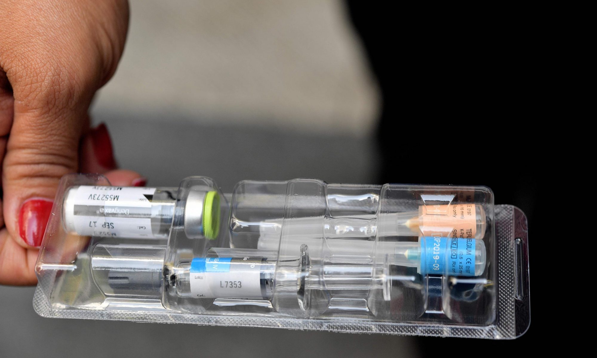
WHO Approves World’s First-Ever Dengue Vaccine

The World Health Organization (WHO) endorsed the world’s first-ever vaccine Dengvaxia for dengue fever. Known as Dengvaxia, the vaccine is the product of two decades of research by French-based Sanofi Pasteur.

To date the vaccine has been approved in Mexico, Philippines, Brazil, El Salvador, Costa Rica, Paraguay, Guatemala, Peru, Indonesia, Thailand and Singapore.

The vaccine is given in three injections spaced out over one year. It is designed for those over the age of nine who have been previously exposed to the virus and is best suited for people living in endemic areas, as opposed to short-term travellers, according to Dr. Alain Bouckanooge, associate vice president of clinical research and development at Sanofi’s division in Thailand.

Studies have shown that overall, the vaccine is effective at reducing dengue by 60 per cent, and reducing severe dengue by 84 per cent.

Against the Den-1 and Den-2 strains – which account for three-quarters of the dengue cases in Singapore – the vaccine’s efficacy is 50 per cent and 40 per cent respectively, compared with 75 and 77 per cent for the other two strains.